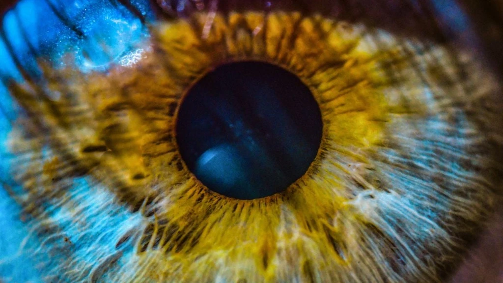
Emerging research confirms the retina serves as a critical window into neurological health. Scientists have established that retinal imaging can now predict Alzheimer’s disease risk years before clinical diagnosis. While these photographs do not diagnose dementia directly, they reveal subtle biological signatures associated with future cognitive decline.

Biomedical engineer Ruogu Fang from the University of Florida led this pivotal study. Fang notes that current diagnostic tools often target late-stage pathology when intervention is no longer viable. Analyzing retinal health offers a novel opportunity to identify at-risk patients early and encourage lifestyle modifications to mitigate disease progression.
The research team deployed machine learning to analyze over 62,000 retinal photographs from UK Biobank participants. Their artificial intelligence model successfully predicted twelve distinct risk factors linked to Alzheimer’s, including vascular stiffening, reduced blood vessel density, and optic nerve thinning. These markers align with broader neurovascular dysfunction observed in dementia patients.
Predictive models integrated demographic, metabolic, and lifestyle data with retinal imagery. Patients who later developed Alzheimer’s exhibited specific constriction of small retinal arteries. This suggests the eye functions as an integrated biological sensor for cumulative systemic risk rather than merely a surrogate questionnaire.
Retinal photography is already standard practice for monitoring diabetes and glaucoma. If validated further, this existing infrastructure could transform routine eye exams into powerful screening tools for brain disease. Published in the Journal of Alzheimer's Disease, this research marks a significant step toward non-invasive, early detection of neurodegenerative conditions.