A pioneering project has revealed the human body like never before, from entire organs down to cellular structures, with unprecedented precision on the scale of a single micron - about 50 times thinner than a strand of hair.
Now, the Human Organ Atlas provides a new gold standard in medical imaging by showing the framework of the brain, heart, lungs, liver, kidneys, and other bodily systems in stunning 3D detail.
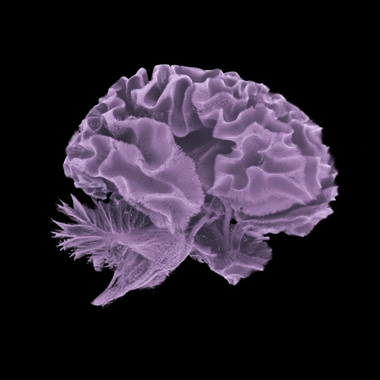
Called hierarchical phase-contrast tomography (HiP-CT), the technique uses X-rays generated by high-energy particles inside a synchrotron called the Extremely Brilliant Source. This accelerator provides a medical imaging tool up to 100 trillion times brighter than regular hospital X-rays.
HOA researchers have non-destructively imaged intact organs from dozens of donors, achieving unique zoom with cellular resolution. The atlas currently includes 87 organs and 363 3D datasets from 54 donors.

HiP-CT has revealed previously unknown disease pathways, including vascular damage in COVID-19 lungs and features of adenomyosis. The atlas also captures cancer and rare pathologies like Dandy-Walker syndrome.
Beyond medical training, the HOA could train machine learning models for improved disease detection. Researchers aim to eventually image complete human bodies with 10 to 20 times higher resolution than currently possible.