A new 5.0 Tesla Susceptibility-Weighted Imaging (SWI) MRI technique shows promise for significantly enhancing the visualization of key brain structures compared to conventional 3.0 T SWI.
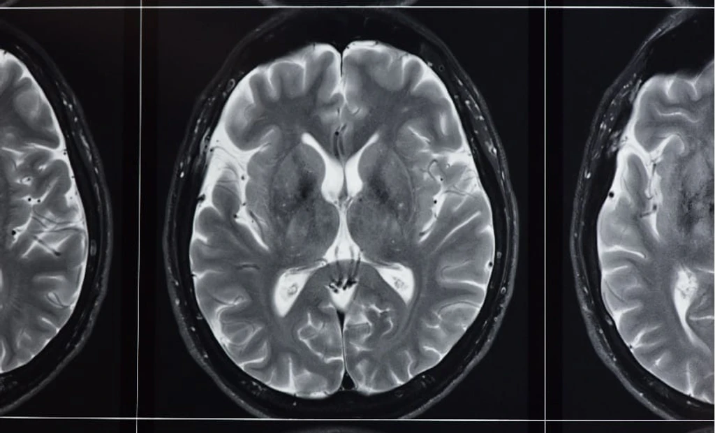
SWI is an MRI method that utilizes magnetic differences between tissues to improve the depiction of cerebral veins and mineral deposits, features crucial in neurodegenerative disease research.
In a comparative study, eight healthy volunteers underwent scans on both 5.0 T and 3.0 T MRI systems. Radiologists found the 5.0 T SWI delivered markedly improved image quality. Contrast-to-noise ratios for cerebral veins increased by over 50%, and more than doubled for deep grey matter structures, indicating substantially enhanced contrast and delineation.
The detection of the 'swallow tail' sign, an imaging feature analyzed in Parkinson's disease, was higher with 5.0 T imaging, identified in 81.25% of cases compared to 50% using 3.0 T.
However, the higher field strength introduced more susceptibility artefacts, particularly near air-tissue interfaces, which can obscure anatomical detail. This highlights an ongoing challenge in high-field MRI: while field strength improves contrast, it can also amplify image distortion.
The findings suggest 5.0 T SWI may improve the clarity of subtle neuroanatomical markers, potentially refining image assessment consistency. Enhanced visualization of cerebral veins and deep grey matter nuclei could support more precise neuroanatomical mapping in studies of neurodegenerative conditions.
Further studies in patient populations are needed to determine if these imaging improvements translate into better diagnostic performance or earlier disease detection.