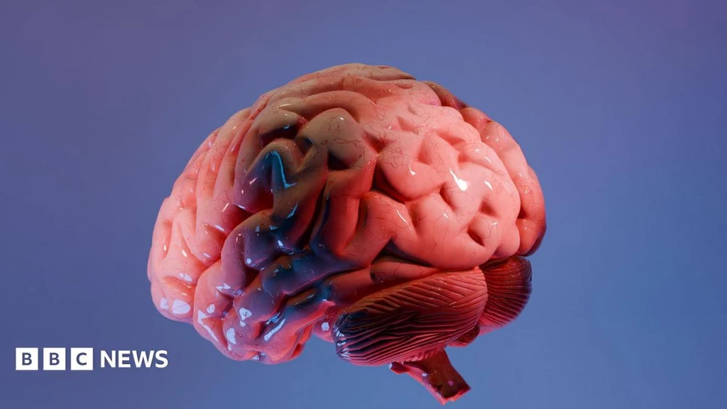
For a century, mapping the human brain has been like charting an unknown continent from scattered observations. Now, scientists at the Sudha Gopalakrishnan Brain Centre at IIT Madras claim a major breakthrough: the world's most detailed three-dimensional atlas of the human brainstem at cellular resolution.
Called Anchor, the atlas combines over 500 tissue sections from foetal, child, and adult brains. It uses high-resolution microscope images and eight chemical markers to create a seamless digital map, allowing users to zoom from an MRI scan of the whole brain down to individual neurons.
The brainstem, though small, is vital. It controls breathing, heartbeat, and movement. Its complex architecture has made detailed mapping impossible-until now.
"We are seeing a visionary programme that puts India at the international table," said Shubha Tole of the Tata Institute of Fundamental Research.
The atlas bridges two separate worlds: medical imaging, which shows the whole brain, and cellular pathology, which examines it one cell at a time. For neuropathologists like Dr. Rebecca Folkerth, who has worked with the team, this is a long-held dream realized.
"What the Indian centre has created is essentially what I dreamed of early in my career," Folkerth said. The current method for diagnosing Alzheimer's involves examining only a tiny fraction of the brain.
The freely available atlas has immediate research potential. By comparing healthy and diseased maps, scientists hope to better understand Parkinson's, stroke, Alzheimer's, and even the long-term neurological effects of COVID-19.
"Detailed brain atlases like this could have a transformative impact," said Partha Mitra of Cold Spring Harbor Laboratory.
The project is part of a broader shift where neuroscience relies heavily on engineering and computation. The team plans to expand the effort, imaging over 100 whole human brains across different ages and diseases to build a comprehensive reference library.