A groundbreaking study suggests that combining PET and MRI scans could accurately distinguish Limbic-predominant age-related transactive response DNA-binding protein 43 kDa encephalopathy (LATE) from Alzheimer's disease (AD).
LATE, characterized by abnormal TDP-43 protein deposits, presents symptoms similar to AD, primarily memory issues, but has different underlying causes and potential treatments. Currently, diagnosing LATE in living patients remains impossible.
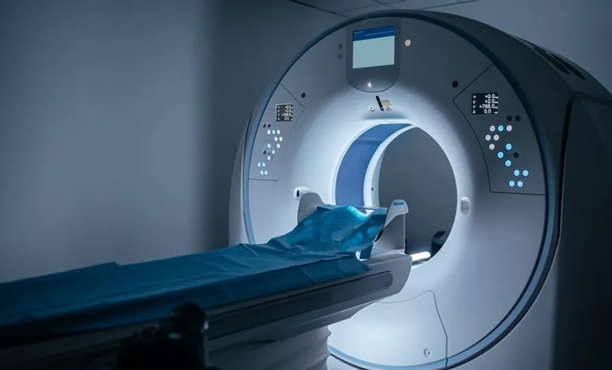
Researchers analyzed nearly 1,000 PET scans and developed advanced imaging templates for both LATE and AD neuropathologic changes. Using autopsy-confirmed data, they categorized participants into probable LATE, mixed LATE and AD, and probable AD.
MRI volumetry findings revealed distinct patterns: the medial temporal lobe was most affected in pure LATE cases, while mixed LATE and AD cases showed vulnerability in the orbitofrontal gyrus and lateral temporal lobe. Key brain regions like the entorhinal cortex and amygdala proved crucial in differentiating these conditions.
This advancement in imaging could pave the way for definitive LATE diagnosis in patients with cognitive impairment, addressing a significant obstacle in understanding and treating this emerging neurodegenerative disorder.