Advanced fetal imaging between 34 and 38 weeks of gestation can predict which newborns with dextro-transposition of the great arteries (d-TGA) will face life-threatening oxygen deprivation after birth.
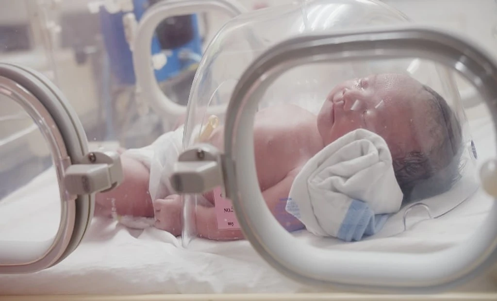
d-TGA is a congenital defect where the aorta and pulmonary artery are reversed, limiting oxygen-rich blood flow. Infants with this condition often require balloon atrial septostomy (BAS) within hours of birth if oxygen saturation drops below 70%.
A prospective cohort study of 34 pregnancies used fetal echocardiography and cardiac MRI to assess risk. Key predictors of urgent BAS included abnormal atrial septal flap morphology, foramen ovale size ≤3.4 mm during maternal oxygen therapy, and altered ductus arteriosus blood flow.
MRI data showing an ascending aorta to pulmonary artery oxygen saturation ratio greater than 1 during maternal hyperoxygenation also strongly correlated with postnatal intervention. The findings suggest combined imaging improves prenatal planning for high-risk cardiac care.